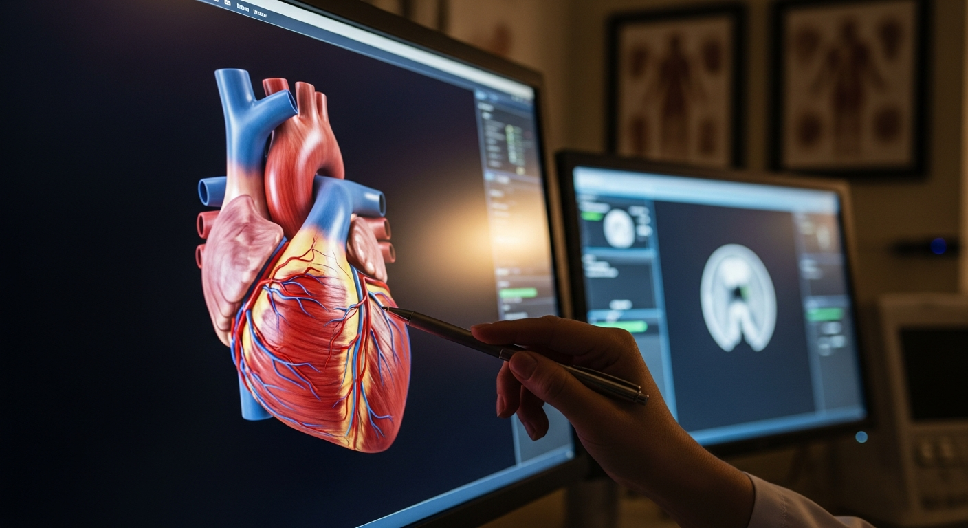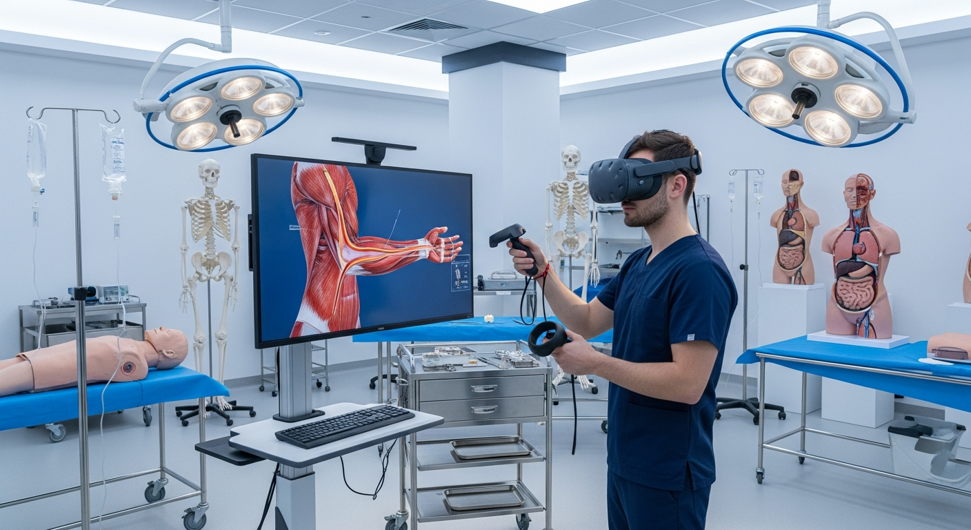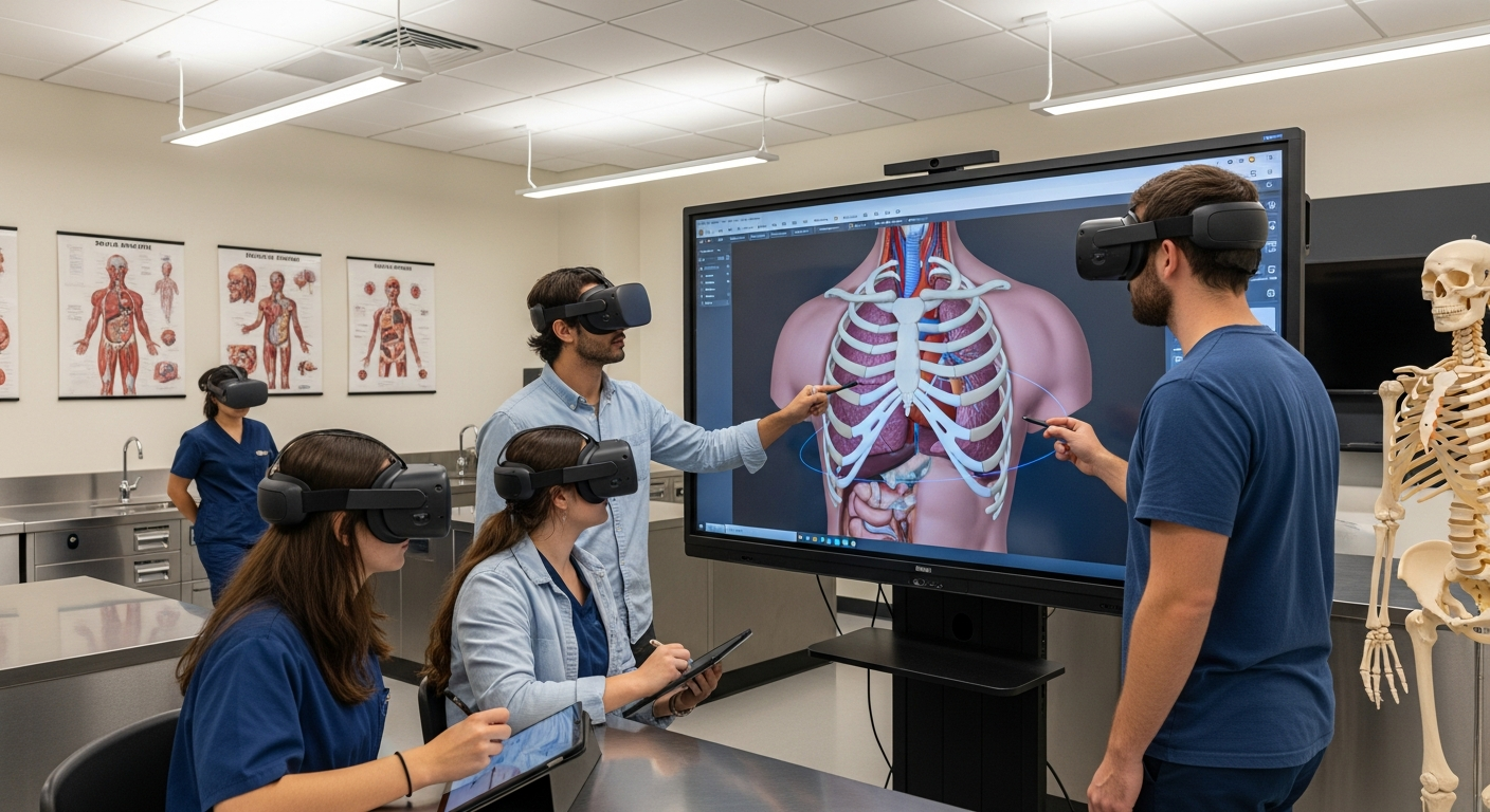
Anatomy education is one of the most essential foundations of medical learning, yet also one of the most historically constrained. For decades, students relied on textbooks, 2D illustrations, limited cadaver access, and static models that could not fully represent the complexity of living anatomy. Extended Reality (XR) — which includes VR, AR, and MR — has transformed that experience. With highly detailed, interactive, and spatially accurate 3D anatomy models, students can now explore the human body with unprecedented clarity and depth.
These XR-powered anatomy models allow learners to rotate organs in 3D space, understand layered structures, practice dissections virtually, and interact with physiological systems in ways that simply aren’t possible with traditional resources. Modern 3D anatomy content is dynamic, intelligent, and immersive — giving medical students, surgeons, and healthcare professionals a more intuitive understanding of human structure and clinical relationships.
This guide explores the advancements in anatomy 3D models through XR, how immersive technology enhances medical knowledge, and why it is rapidly becoming the standard in healthcare learning and simulation.
1. How XR Elevates Anatomy Education Beyond Traditional Methods?
Traditional anatomy teaching depends heavily on cadavers, textbooks, and 2D diagrams. These methods have limitations:
limited access to cadavers
structural degradation over time
inability to show real function
static images that don’t convey perspective
lack of safe repeat practice
XR solves these problems by enabling anatomy exploration inside immersive digital environments, similar to the improvements seen in VR medical training methods, reshaping clinical education.
XR 3D anatomy models allow students to:
zoom in and out of organs
peel back layers of tissue
isolate systems (muscular, skeletal, vascular, etc.)
explore structures from any angle
simulate functional movement
repeat dissections without limitation
This multi-layered, repeatable exploration offers a richer understanding than any static model.
2. High-Fidelity 3D Models With Realistic Detail
Recent advancements in 3D modeling and medical imaging have greatly improved the realism of digital anatomy.
Modern XR anatomy models feature:
ultra-high-resolution textures
accurate spatial proportions
micro-anatomical detail (e.g., nerve branches, capillary patterns)
dynamic lighting and shadows
lifelike color, shading, and movement
These improvements build on innovations introduced in 3D medical training innovations, where digital models capture medical precision at scale.
Why realism matters:
supports surgical training
enhances diagnostic understanding
improves spatial recall
prepares learners for real procedures
The closer digital anatomy gets to real-life structures, the more effective XR training becomes.
3. XR Enables Layer-by-Layer Anatomy Exploration
One of XR’s strongest educational benefits is the ability to explore anatomy layer by layer — something no physical model can fully replicate.