Surgical training has always demanded precision, practice, and exposure to real-world variation — yet traditional learning pathways often limit how often trainees can rehearse procedures or experience high-risk situations. Cadaver availability, OR access, time constraints, and patient safety concerns all restrict the consistency and volume of hands-on practice medical teams receive. Extended Reality (XR) changes this entirely.
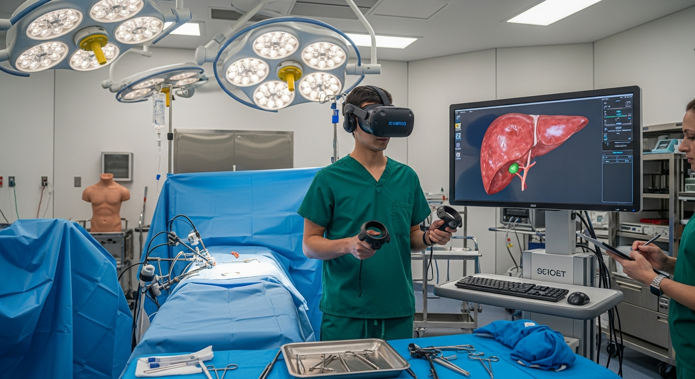
Through VR, AR, and MR systems, surgical simulation has evolved into an advanced training environment where learners can practice complex procedures repeatedly, safely, and with a level of anatomical accuracy that closely mirrors real patients. These simulations use intelligent overlays, real-time feedback, digital twins, and anatomy-specific visualization to prepare clinicians for the operating room long before they ever enter it.
This guide explores how XR surgical simulation works, why it has become the new standard for procedure-ready training, and how immersive technologies elevate surgical competency across the healthcare ecosystem.
1. Why XR Surgical Simulation Is Transforming Modern Training
XR introduces a level of repeatability, safety, and visualization that traditional surgical training struggles to match. In VR, students can practice procedures dozens of times without risk. In AR and MR, clinicians receive digital guidance layered onto real tools or mannequins, enhancing accuracy and decision-making.
This immersive approach strengthens foundations previously built through VR medical training workflows, where repetition and realism dramatically improve readiness.
Key advantages include:
unlimited procedural repetition
high-stakes practice without patient risk
detailed anatomy exploration
Reduced anxiety entering real OR settings
measurable performance scoring
XR ensures learners gain confidence and skill before their first real patient encounter.
2. Anatomy-Accurate, High-Fidelity Surgical Environments
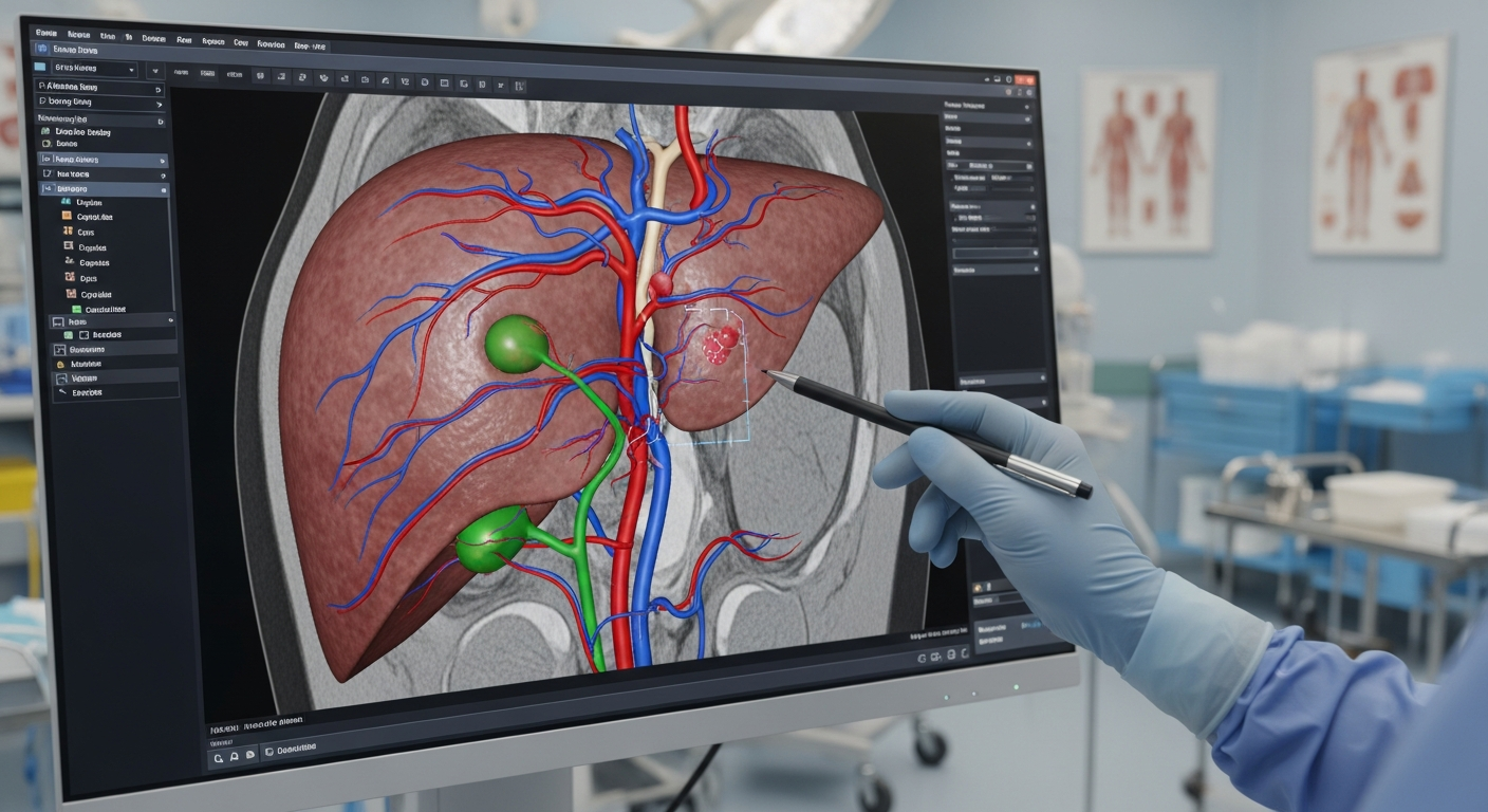
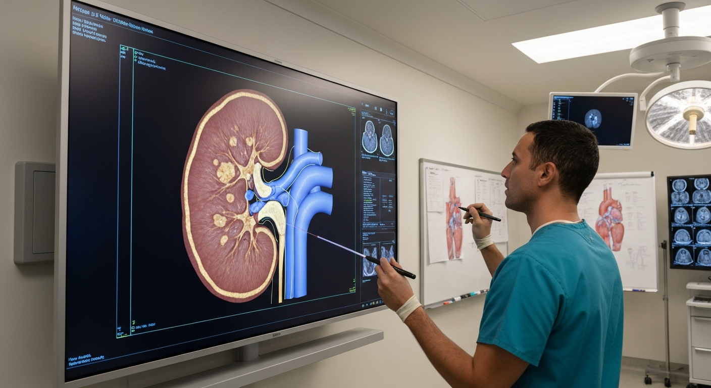
Modern surgical simulations replicate real human anatomy with incredible precision.These lifelike models are powered by 3D reconstruction, volumetric imaging, and detailed texture mapping.
The sophistication of these models builds on 3D medical training innovations, where anatomy accuracy directly influences clinical competency.
XR surgical environments include:
full-organ systems with layered structures
realistic tissue responses
accurate instrument physics
pathology-specific variations
dynamic bleeding and suction behavior
Trainees learn not just the ideal procedure — but the anatomical diversity they will encounter in real hospitals.
3. Procedure Walkthroughs With Step-by-Step XR Guidance
XR offers guided surgical workflows that mirror expert-level procedures.These guides appear as intuitive overlays, tool paths, alerts, and contextual prompts.
Future-ready XR guidance will include:
incision lines placed directly on the patient model
holographic instruments showing motion arcs
risk zone visualization (nerves, arteries, critical landmarks)
dynamic adjustments based on user behavior
AI-generated suggestions for next steps